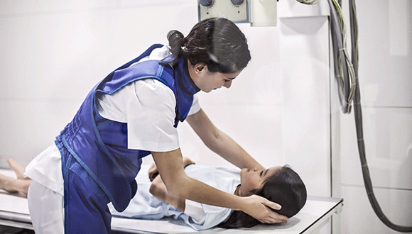
Should you limit your X-ray exposure?
X-rays have been used in medicine for more than 120 years. What risks do we need to be aware of?
Dr Lindy Alexander
July 2018
X-rays use a type of radiation called electromagnetic waves to create pictures of the inside of your body. The X-rays go through soft structures like skin easily but slow down through dense structures like bone, so these show up as white on the image.
They don’t only spot broken bones; different types of X-rays can also identify pneumonia, breast cancer, tooth decay and digestive problems.
CT scans, introduced in the 1970s, use many X-rays at different angles to get a more detailed picture inside the body. They can be used to find varied health problems, from kidney stones to tumours, and can also help in guiding treatment and surgery.

Are X-rays dangerous?
We’re naturally exposed to radiation every day through the environment – the sun, stars, air and earth are all sources of radiation.
High levels of radiation can damage your cells, which could lead to health issues like cancer. However, as X-rays have relatively low radiation, the risks associated are minimal.
The level of radiation from an X-ray depends on the part of the body being scanned. A chest X-ray has about the same level of radiation as 2.4 days of environmental radiation. A CT scan can have up to 200 times more radiation. Speak to your doctor about whether the risks outweigh the benefits.
The first X-ray – of the hand of a young woman – was taken in 1895. It was a huge breakthrough in medicine but subjected the patient to a radiation dose 1,500 times higher than today’s X-rays.
Can X-rays harm my children?
Because children’s bodies are still growing and developing, they’re more sensitive to the risks of radiation. If you could be pregnant, discuss the risks with your doctor, particularly if you need to have an X-ray of the abdominal area. Where possible, children should have an alternative first, such as an ultrasound.
Related articles
HOW CT SCANS WORK
Professor John Slavotinek, Dean of Clinical Radiology at the Royal Australia and New Zealand College of Radiologists explains.
HOW MRIS WORK
Professor John Slavotinek, Dean of Clinical Radiology at the Royal Australia and New Zealand College of Radiologists explains.
HEALTH CHECKS BY AGE: THE TESTS YOU SHOULD BE HAVING
Your guide to staying on top of your health, through every stage of life.
PERSONALISING BREAST CANCER TREATMENT FOR BETTER RESULTS
Future breast cancer treatments are evolving towards targeted therapies individualised for each patient’s condition.